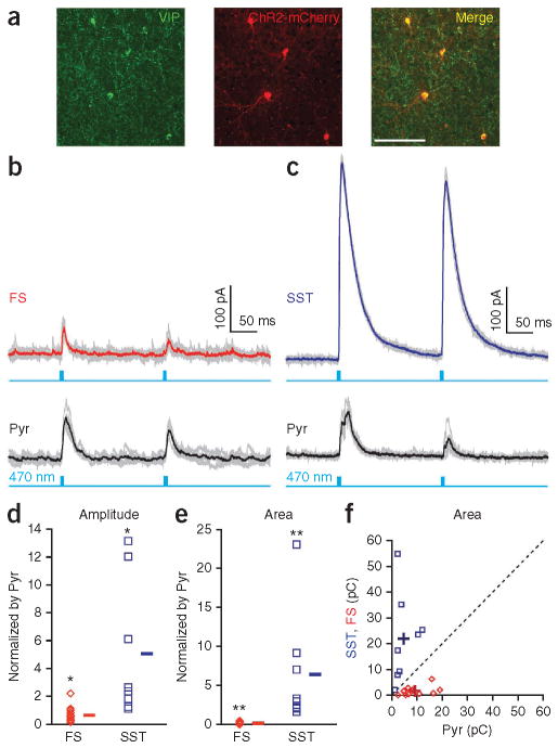
Figure 3.
VIP interneurons most strongly inhibit SST interneurons in S1 superficial layers. (a) Specific expression of ChR2-mCherry in VIP interneurons. ChR2-mCherry expression in Vip-cre mice was confined to VIP neurons. Antibody-stained VIP neurons (left) showed nearly 100% overlap (right) with neurons expressing ChR2-mCherry (middle). Scale bar represents 100 μm. (b) VIP interneurons provided weak inhibition to fast-spiking interneurons. Photo-stimulation–evoked inhibitory synaptic currents recorded in a fast-spiking interneuron and in a pyramidal neuron in the Vip-cre B13 mouse are shown. (c) VIP interneurons provided strong inhibition to SST interneurons. Photo-stimulation–evoked inhibitory synaptic currents in a SST interneuron and a simultaneously recorded pyramidal neuron in a Vip-cre GIN mouse are shown. Cesium-based internal pipette solution was used to record inhibitory currents at 0 mV in voltage-clamp mode. Gray traces indicate individual sweeps and colored traces indicate the average (red, fast spiking; blue, SST; black, pyramidal neurons). Light blue traces indicate photo-stimulation (LED, 470 nm, 5 ms) delivered at 5 Hz. (d,e) IPSC amplitude (d) and total charge (e) in fast-spiking (14 cells, 8 slices, 4 mice) and SST (8 cells, 5 slices, 3 mice) interneurons normalized to the corresponding values in simultaneously recorded nearby pyramidal neurons. *P < 0.05, **P < 0.005, Wilcoxon signed-rank test. (f) Photo-stimulation–evoked total IPSC charge in fast-spiking interneurons (red) and SST interneurons (blue) plotted against total IPSC charge in simultaneously recorded pyramidal neurons. Dashed line indicates unity. + indicates mean (fast spiking to pyramidal ratio: amplitude, P = 0.02; charge, P < 0.001; SST to pyramidal ratio: amplitude, P = 0.01; charge, P < 0.001).

