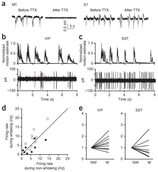
Figure 6.
vM1 activity is responsible for the increased activity of VIP interneurons and decreased activity of SST interneurons in S1 during whisking. (a) TTX injection to vM1 silenced the activity of vM1, but did not affect S1 activity, as evidenced by LFPs recorded in S1 and vM1 before and after TTX injection to vM1. TTX injection and LFP recordings were conducted under anesthesia. (b,c) Representative recordings from a VIP interneuron (b) and an SST interneuron (c) during vM1 inactivation. Whisking motion was computed from whisker video recordings to define whisking (W) and non-whisking (NW) periods (top). Bottom, spikes in the corresponding interneuron type recorded under two-photon guidance in loose-patch configuration. (d) Firing rates of VIP (open circles) and SST (black circles) interneurons during whisking plotted against their firing rates during non-whisking periods after vM1 inactivation. Dashed line indicates unity. (e) Summary data comparing firing rates of VIP (9 cells, 2 mice) and SST (8 cells, 3 mice) interneurons during whisking and non-whisking periods after inactivation of vM1 with TTX injection (VIP interneurons, P = 0.08; SST interneurons, P = 0.2, Wilcoxon signed-rank test).

