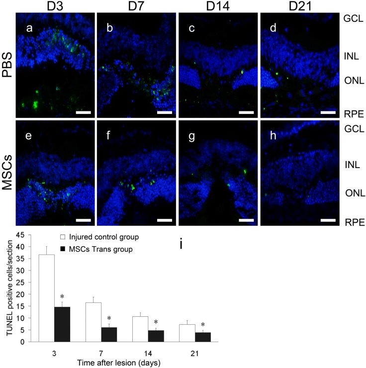
Figure 3.
Apoptosis of retinal cells at laser spots in mice treated with or without MSCs. Retinas of the injured control group (a–d) and MSC-treated group (e–h). Green indicates terminal deoxynucleotidyl transferase-mediated dUTP-biotin nick-end labeling (TUNEL)+ cells. Blue indicates DAPI-stained nuclei. Scale bars: 50 μm. The average numbers of apoptotic cells were calculated and found to be significantly reduced in the MSC-treated group at all time points compared with those in the injured control group (i). *, p < 0.05. Scale bars: 50 µm. GCL, ganglion cell layer; INL, inner nuclear layer.

