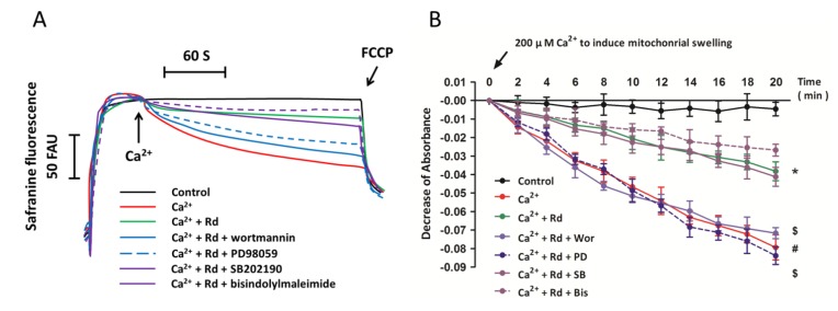
Figure 6.
Effects of protein kinase inhibitors on mitochondrial membrane potential dissipation and swelling after Ca2+ treatment in Rd pretreated spinal cord mitochondria. Isolated spinal cord mitochondria (0.5 mg protein/mL supported by succinate) were pretreated with 1 μM wortmannin (Wor, PI3K inhibitor), 50 nM bisindolylmaleimide (Bis, PKC inhibitor), 10 μM PD98059 (PD, ERK inhibitor), or 10 μM SB202190 (SB, p38 inhibitor) in the presence or absence of 10 μM Rd for 10 min before the incubation with 30 μM Ca2+. (A) The mitochondrial membrane potential was measured up to 300 s. Controls were performed in the absence of Ca2+, and 1 μM FCCP was added to induce maximal depolarization at the end of the measurements. Traces are representative of three independent experiments performed in duplicate and were expressed as fluorescence arbitrary units (FAU); (B) Mitochondrial swelling was examined by monitoring the absorbance at 540 nm induced by 200 μM Ca2+. The control was measured without Ca2+. The data were represented as means ± SD from five experiments. # p < 0.05 vs. control. * p < 0.05 vs. Ca2+. $ p < 0.05 vs. Ca2+ + Rd.

