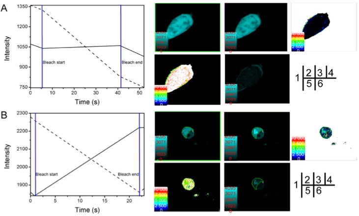
Figure 3.
Verification of SelP' and α-tubulin interaction by the receptor photobleaching method of FRET. HEK293T cells were co-transfected with the empty plasmids pECFP-C1 and pEYFP-C1 as a negative control (A) or co-transfected with pECFP-C1-SelP' and pEYFP-C1-Tub for sample tests (B). (1) Photobleaching curves (solid lines for donor fluorescence and dashed lines for receptor fluorescence). The region of interest (ROI) was bleached at 515 nm; (2) The fluorescence images of donors before bleaching; (3) The fluorescence images of donors after bleaching; (4) Donor fluorescence increments before and after bleaching; (5) Diagram of the distance between donor and receptor; (6) FRET efficiency diagram.

