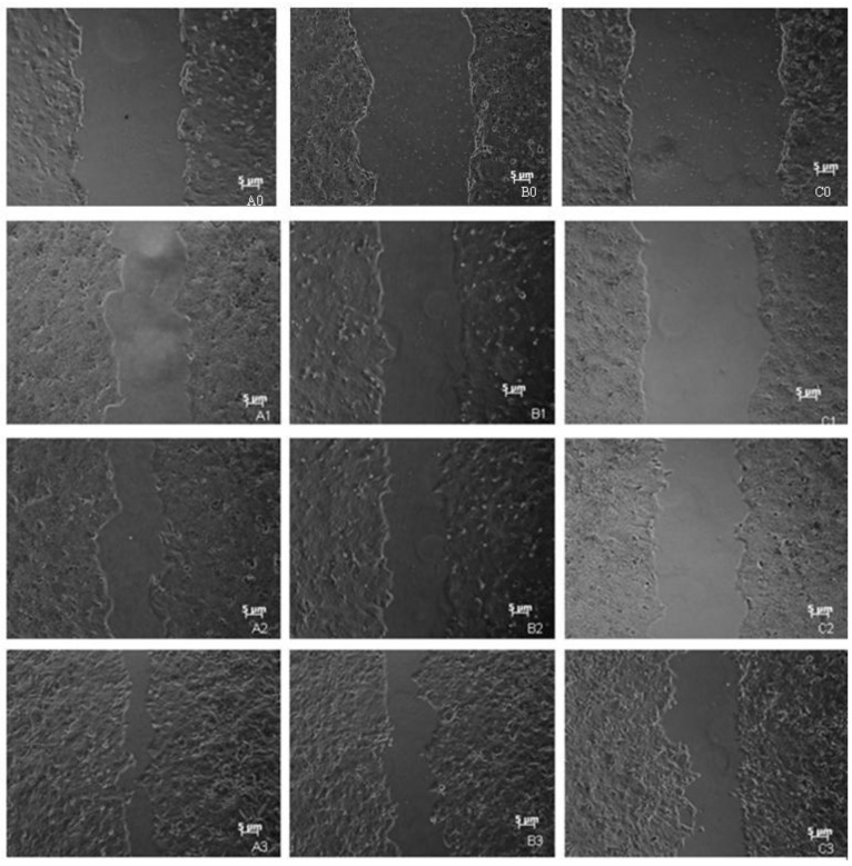
Figure 3.
Wound distance analysis via scratch wound assays. A0, A1, A2, A3: images of control cells incubated for 0, 24, 48, and 72 h (50×); B0, B1, B2, B3: images of EGFP-C1 recombinant plasmid transfected cells incubated for 0, 24, 48 and 72 h (50×); C0, C1, C2, C3: images of EGFP-C1-HSPC117 recombinant plasmid transfected cells incubated for 0, 24, 48, and 72 h (50×).

