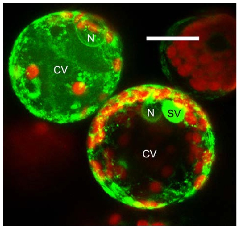
Figure 1.
In this 10 μm projection of confocal images, two tobacco protoplasts over-expressing the Green Gluorescent Protein harboring the C-terminal Vacuolar Sorting Determinant of chitinase A (GFPChi, in green) accumulate the transgene following the two most described patterns. In the upper cell the transgene labels the endoplasmic reticulum (as the nuclear envelope is visible) but efficiently reaches the central vacuole. Several smaller compartments are also visible; In the lower cell the transgene also labels the ER but is not accumulated in the central vacuole. On the other hand, it is strongly accumulated in small vacuoles [8]. The red fluorescence of chlorophyll labels chloroplasts. N, nucleus; CV, central vacuole; SV, small vacuole. Scale bar = 20 μm.

