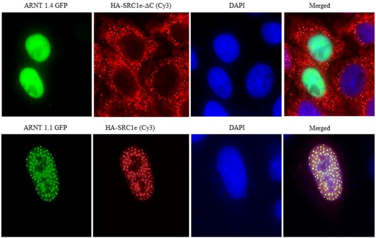
Figure 2.
HeLa cells after cotransfection with the indicated hARNT1.1, hARNT1.4, SRC1e, and SRC1e-ΔC constructs. Green indicates GFP, red indicates HA-CY3 staining, and blue indicates nuclear DAPI staining (adapted from [23]). Twenty-four hours after transfection, fixed cells were incubated with HA antibodies (12CA5 hybridomas, MBL) for 1 h, washed, and then incubated with Cyanine 3 (Cy3)-conjugated secondary antibodies for 30 min. The cells were then washed again and mounted in Vectashield (Vector Laboratories, Burlingame, CA, USA) mounting medium containing 4,6-diamidino-2-phenylindole (DAPI). Fluorescence images were visualized using an Olympus 1670 inverted system microscope (Olympus Optical Co., Ltd, Tokyo, Japan) equipped with a charge-coupled device (Magnification ×1000).

