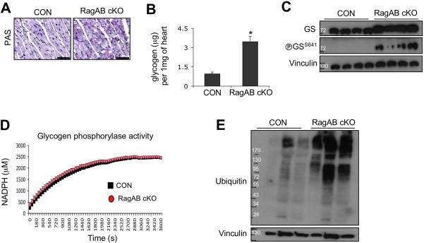
Figure 10. Cardiac hypertrophy in RagA/B cKO mice exhibits the features of lysosomal storage diseases.
(A) Periodic acid-Schiff staining of heart tissues. Representative images are shown. Scale bars, 50 μm. (B) Biochemical measurement of glycogen level in heart tissues. Glycogen levels were determined using an enzyme-based colorimetric assay kit. Values represent the mean ± SD of data (n=4 per group). *P < 0.03, Wilcoxon rank sum test. (C) Immunoblot analysis of glycogen synthase-1 in heart. Tissue lysates were prepared from control and RagA/B cKO hearts, and levels of glycogen synthase and phosphoglycogen synthase were investigated by immunoblotting. (D) Glycogen phosphorylase activity assay. Using an enzyme-based assay, activity of glycogen phosphorylase in heart tissue was determined by measuring NADPH production in the reaction. Each point represents the mean ± SD of data (n=4 per group). (E) Accumulation of ubiquitinated proteins in RagA/B cKO hearts. Levels of ubiquitinated proteins in heart tissues were examined by immunoblotting.

