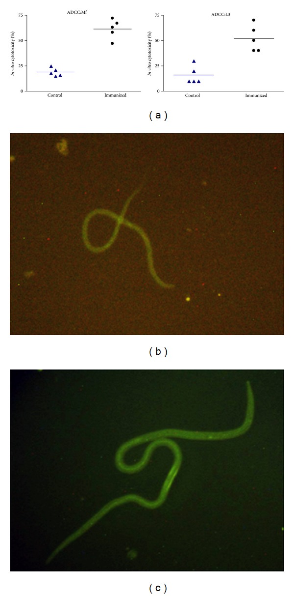
Figure 10.

Antibody dependent cellular adhesion to Mf and L3 of B. malayi. Ten L3 and 100 Mf were taken per well and were incubated with PEC isolated from normal Mastomys in the presence of sera from Bm-iPGM immunized animals. (a) Sera of Bm-iPGM immunized mice promoted adherence of PEC to Mf and L3 larvae and induced significant death of Mf (61.40% cytotoxicity) and L3s (52%). Photographs were captured on phase contrast microscope (Nikon, Japan) at 40x magnification. Data are presented as mean ± S.E. values from five different wells. Interaction of anti-Bm-iPGM antibodies with B. malayi Mf (b) and L3 (c) as shown by fluorescence microscopy. Parasites were incubated with anti-Bm-iPGM sera for 4 h and further incubated with FITC labelled anti-mouse IgG for 2 h. Images were captured under fluorescent microscope at 20X for Mf and 10X for L3.
