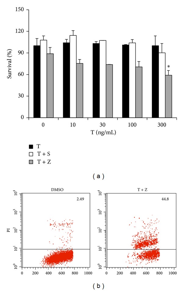
Figure 2.

HT-22 hippocampal neurons are committed to necrosis rather than apoptosis in response to TNF-α. (a) HT-22 hippocampal neuronal cells were treated as indicated for 20 h. Cell viability was determined by measuring ATP levels. Data are represented as mean ± standard deviation of duplicates. T: TNF-α; S: Smac mimetic (100 nM); and Z: z-VAD (20μM). (b) HT-22 hippocampal neurons were treated with DMSO or TNF-α (300 ng/mL)/z-VAD for 20 h and then analyzed for PI staining by flow cytometry. Identical concentrations were used in later experiments. Data are represented as mean ± standard deviation of duplicates. *P < 0.01, **P < 0.001 versus control. All experiments were repeated three times with similar results.
