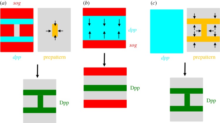
Figure 3.
Schematic diagram of directional transport of Dpp/BMP ligands. (a) Top left: complementary expressions of dpp (light blue) and sog (red). Top right: prepatterned information (orange) that instructs the direction of Dpp transport. Bottom: Dpp proteins move to the prepatterned position (green), e.g. PCV in Drosophila. (b) Top: complementary expression of dpp (light blue) and sog (red). Sog diffusion instructs the direction of Dpp movement. Bottom: Dpp is redistributed to the midline (green), e.g. early embryo in Drosophila. (c) Top left: ubiquitous expression of dpp mRNA (light blue). Top right: prepatterned information (orange) that instructs the direction of Dpp transport. Bottom: Dpp is redistributed to the prepatterned position (green), e.g. wing vein formation in the sawfly.

