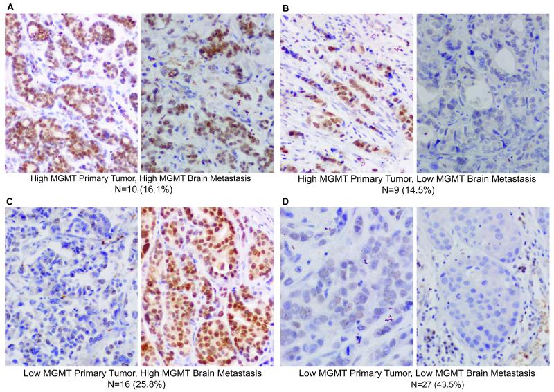
Figure 4. MGMT expression in matched sets of human breast primary tumors and resected brain metastases.
A-D. Sixty-two patient matched sets were collected from tumor banks in Poland and Germany. TMAs of the specimens were stained for MGMT and evaluated for the percent of positively staining tumor cells (nuclear staining only), dichotomized at 5%. The number and percentage of specimens in each category is given below each representative photomicrograph.

