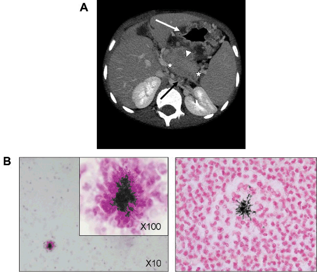
Figure 1.
A, Radiologic signs of abdominal actinomycosis on a computed tomographic (CT) scan (patient 6). Abdominal ultrasonography revealed segmentary portal hypertension and perigastric and esophageal varices (not shown); CT scan showed splenomegaly and thrombosis of the splenic vein (extension delimited by white stars), multiple mesenteric lymph nodes (black arrow), gastric wall thickening (white arrow), and a peripancreatic mass (white arrowhead). B, Histologic analysis of abdominal actinomycosis (patient 6). Liver histologic analysis after liver puncture in patient 6 showed extensive parenchymal necrosis and multiple abscesses with filamentous gram-positive bacteria within the granule surrounded by neutrophil “clubbing” cells typical for actinomycosis (original magnification, x10 and x100; Gram stain on the left and Grocott stain on the right).

