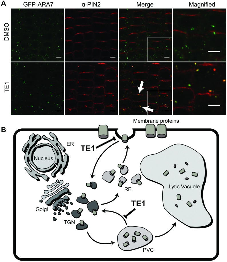
Figure 4. TE1 causes protein accumulation at the PVC.
Seedlings expressing GFP–ARA7 were incubated in DMSO (upper row) and 25 μM TE1 (lower row) for 120 min. The first column shows GFP–ARA7 and the second column shows immunolabelling of PIN2. The last two columns are merged images of GFP–ARA7 (the last column is a magnification of a section of the third column) and labelling with anti-PIN2 antibody (A). Scale bars, 5 μm. (B) Endomembrane protein trafficking and possible mode of action of TE1.

