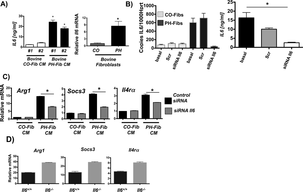
Figure 4. PH-Fibs activate macrophages through paracrine IL6.
(A) IL6 protein amounts in CM from 2 bovine PH Fib populations; mean +/− SEM are shown from analysis of triplicate samples of each and compared to IL6 amounts in CM from 2 CO-Fibs, also tested in triplicate; * depicts p< 0.05 by t-test (PH-Fib CM vs. Cntrl CM); and relative IL6 mRNA in bovine CO-Fib (normalized to 1, n=5) vs. PH-Fib (n=4); * depicts p< 0.05 by t-test. (B) siRNA mediated knock down of IL6 transcription in bovine CO-Fibs and PH-Fibs; Scr=scrambled, basal=untreated; and IL6 protein amounts in CM from untreated (basal), scrambled, and IL6 siRNA treated PH-Fibs (n=3 each) tested in triplicate ELISA assay. (C) siRNA-mediated suppression of IL6 gene transcription in bovine PH-Fibs limits the ability of CM to induce transcription of STAT3 regulated genes in WT mouse BMDMs. Gene expression is normalized to expression of Hprt1 and relative to that in macrophages exposed to CM from CO-Fibs treated with control siRNA. Data are mean ± SEM from triplicate determinations and representative of two separate experiments. (D) Expression of STAT3 regulated genes, including Hif1α, in WT and Il6−/− BMDMs exposed for 16 hrs to bovine PH-Fib CM. *P < 0.05 by unpaired two-tailed Student’s t-test of triplicate PCR analysis; one representative experiment with CM from one of three PH-Fib populations was tested on BMDMs from 3 different animals of each genotype.

