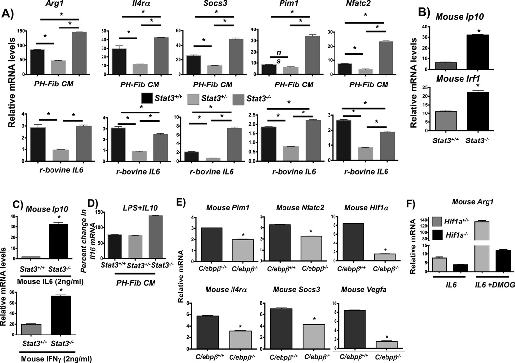
Figure 8. STAT3, C/EBPb, and HIF1a are critical regulators of fibroblast-mediated macrophage activation.
(A) Transcription of Arg1, Il4ra, Socs3, Pim1, and Nfatc2 in BMDMs from Stat3fl/flTie2cre (designated Stat3−/−) mice compared to BMDMs from WT mice (Stat3+/+) and from Stat3fl/+Tie2cre (designated Stat3+/−) in response to bovine PH-Fib CM and recombinant bovine IL6 (2ng/ml) after stimulation for 16 hrs. (B) STAT1 target gene expression (Ip10 and Irf1) in Stat3−/− compared to WT BMDMs in response to bovine PH-Fib CM after stimulation for 16 hrs. (C) STAT1 target gene expression (Ip10) in response to stimulation with mouse recombinant IL6 (top panel), or mouse recombinant IFNγ (bottom panel) in Stat3−/− compared to WT BMDMs. (D) Percent gene expression of Il1b in WT, Stat3+/−, and Stat3−/− BMDMs in response to LPS (100ng/ml)+IL10 (10ng/ml) compared to LPS (100ng/ml) alone (set as 100%) after 16 hrs of incubation. (E) Gene expression of Pim1, Nfatc2, Hif1a, Il4ra, Socs3, and Vegfa in C/ebpb−/− mouse BMDMs compared to WT BMDM in response to bovine PH-Fib CM after 16hrs of stimulation. (F) Expression of Arg1 in LysMcre (designated Hif1a+/+) and Hif1afl/flLysMcre (designated Hif1a−/−) mouse BMDMs in response to mouse recombinant IL6 (after stimulation for 16hrs) in the presence or absence of HIF stabilization with DMOG. Data are obtained from PCR triplicates and representative of results obtained from at least two different PH-Fib isolates and BMDMs from 2 different animals. *P < 0.05 by unpaired two-tailed Student’s t-test. *P < 0.05 by unpaired two-tailed Student’s t-test.

