
Figure 1.
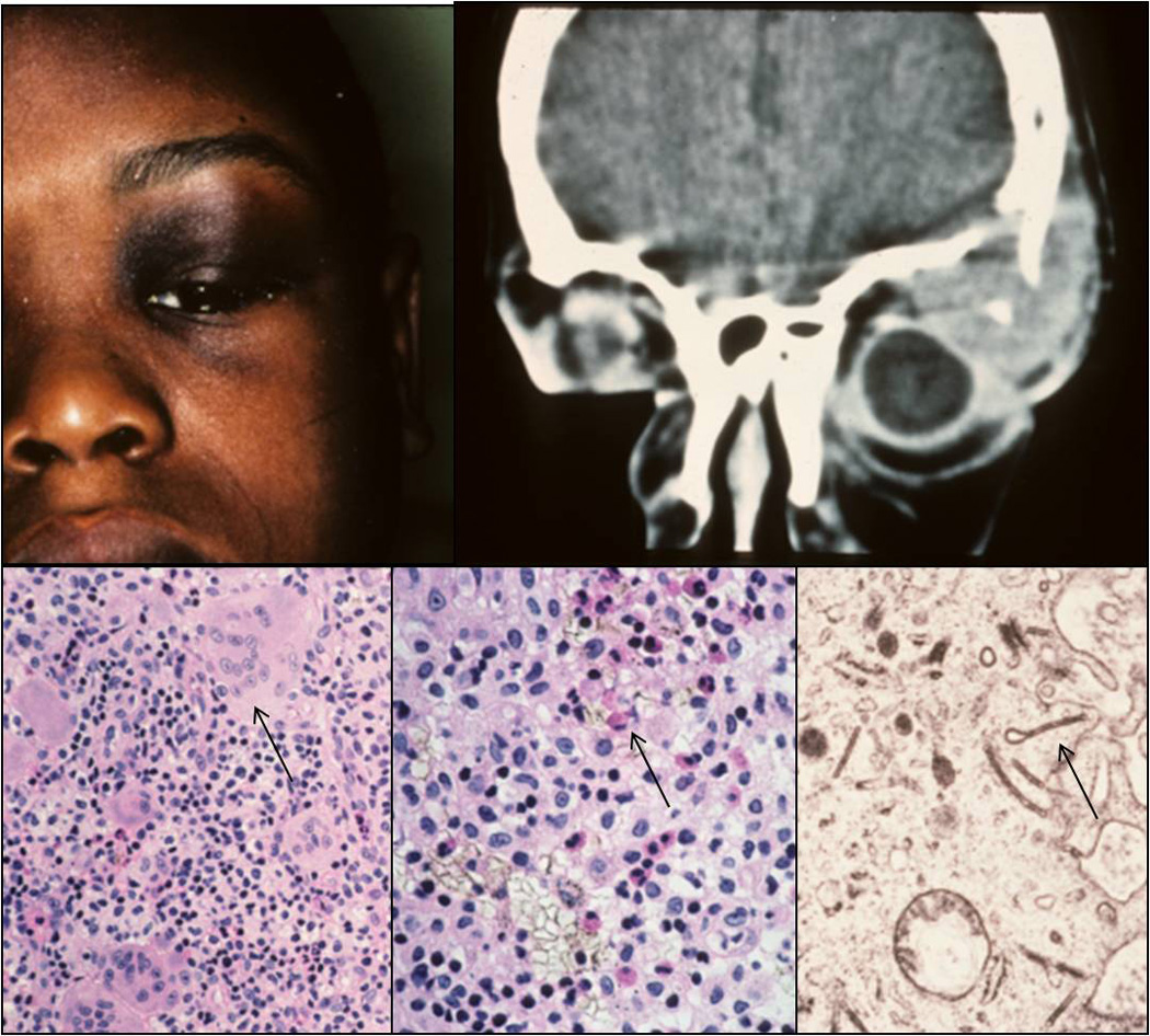
Clinical appearance of an 8-years-old patient with a superior temporal mass in the left orbit (A). Computed tomography (CT) demonstrating the orbital lesion with bone erosion (B). Histology of the lesion showing infiltrates composed of histiocytes, giant cells (C, arrow; H&E, 80x), eosinophils (D, arrow; H&E, 128x), and lymphocytes. Birbeck granules exhibiting the shape of tennis rackets were detected by transmission electron microscopy (E; TEM, 30000x).