
Figure 3.
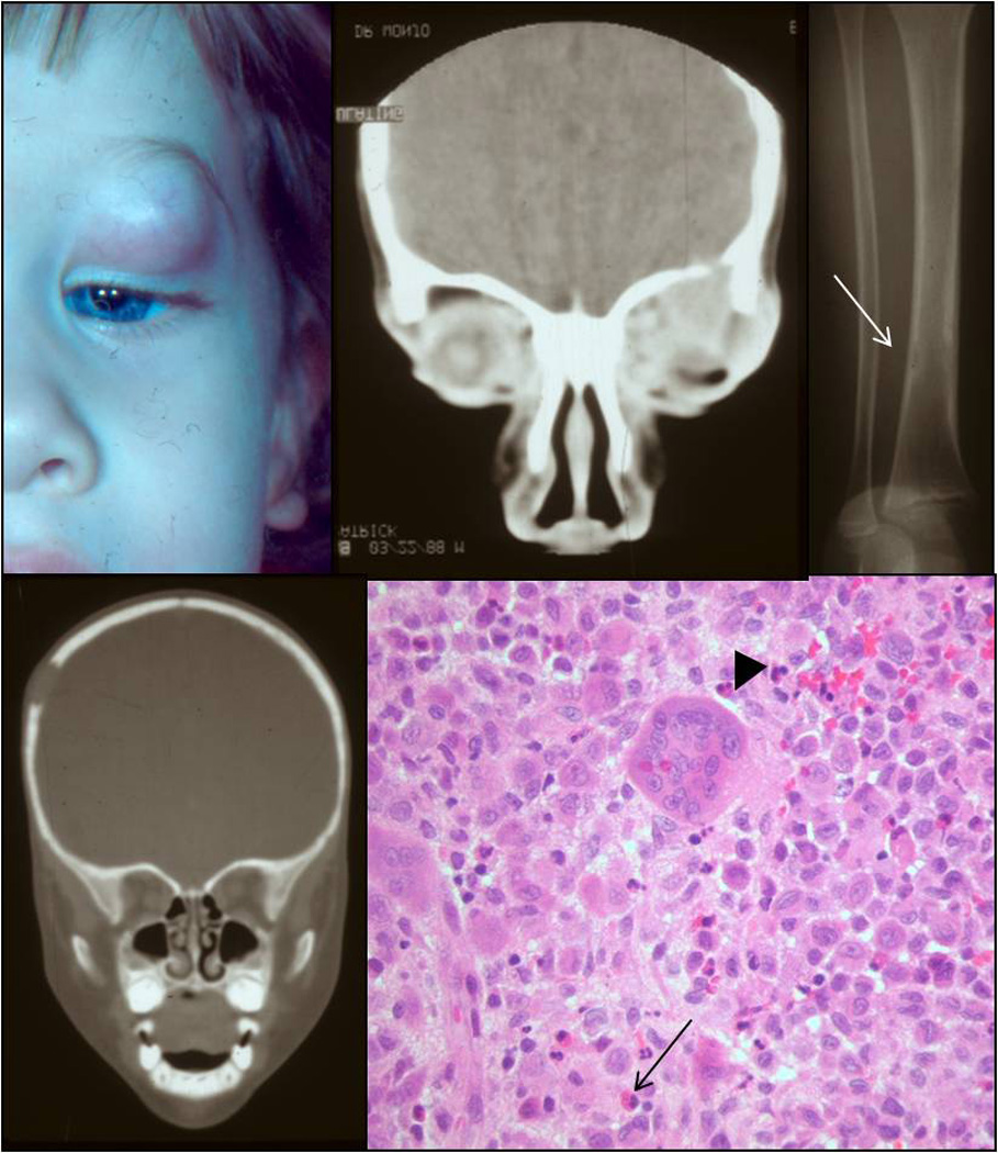
Clinical appearance of a one-years-old patient with a mass in the anterior orbit (A). CT revealing bony erosion in the orbital rim (B), the tibia (C), and the skull (D). Histology showing sheets of histiocytes intermixed with giant cells (arrowhead) and eosinophils (arrow) (E; H&E, 40x).