
Figure 5.
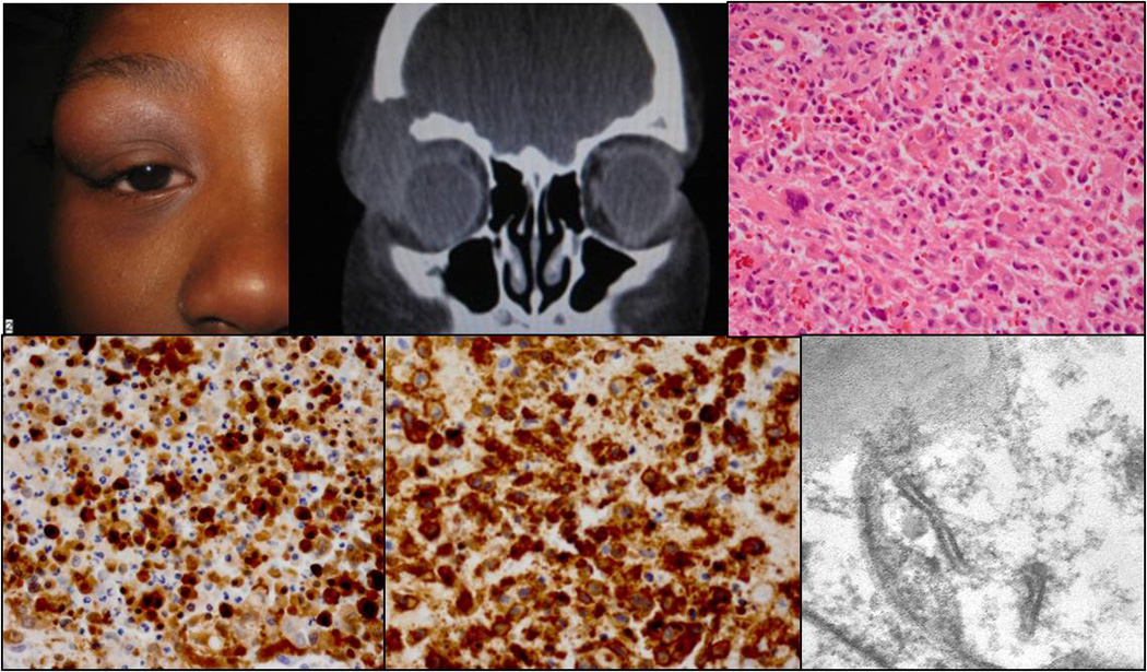
Clinical appearance of a nine-years-old patient with a mass in the right upper orbit (A). CT demonstrating the orbital lesion with bone erosion (B). Histology illustrating the Langerhans cell infiltrate with giant cells and eosinophils (C; H&E, 40x). The Langerhans cells were positive for S100 (D; 40x) and CD1a (E; 40x). Birbeck granules were detected by TEM (F; 14000x).