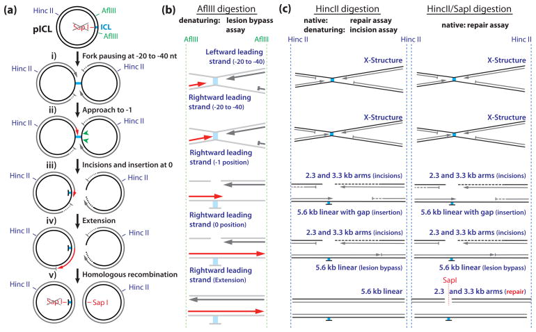
Fig 1.
Schematic Overview of DNA repair intermediates and products generated by the various assays described in this chapter. (a) Cartoon of pICL showing the restriction sites and intermediates formed during repair. (b) DNA products analyzed on sequencing gels in the lesion bypass assay (Subheading 3.3). Dark grey and red strands are visible on the gel in Fig. 2. Lesion bypass of the rightward moving fork (red strands) can be followed at single nucleotide resolution (see Fig. 2). (c) Products analyzed under denaturing and native conditions in the incision and repair assays respectively (Subheadings 3.4 and 3.5).

