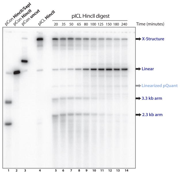
Fig. 3.
Examination of dual incisions during ICL repair. HincII digested repair intermediates are separated on a denaturing gel and visualized using Southern blotting (land 5–14) (Subheading 3.4). Undigested and HincII and/or SapI digested pControl and pICL are shown in lane 1–4 and serve as size markers. X-structures, linears, and 2.3/3.3 arm fragments are indicated. pQuant is visible due to some background reactivity with the probe.

