Figure 1.

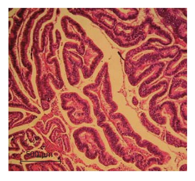
Hematoxylin and Eosin staining of the liver specimen (10 × magnification). The histopathological analysis of the specimen indicates a well-differentiated peripheral cholangio-carcinoma with no vascular invasion.

Hematoxylin and Eosin staining of the liver specimen (10 × magnification). The histopathological analysis of the specimen indicates a well-differentiated peripheral cholangio-carcinoma with no vascular invasion.