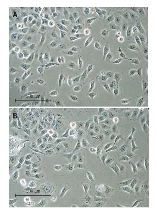
Figure 2.

The RMCCA-1 culture under a phase contrast microscope at 20 × magnification. (A) at 16th passage; (B) at 30th passage. The RMCCA-1 cells exhibited circular to spindle shape with many processes and ornamental fringes. The nucleus and cytoplasm appeared granulated.
