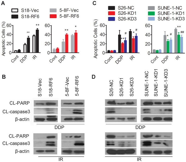
Figure 4. Upon cisplatin or radiation treatment, RASSF6 overexpression increased apoptosis, and RASSF6 depletion reduced apoptosis.
(A, B) S18 and 5-8F cells with stable RASSF6 overexpression (RF6) or transfected with an empty vector control (Vec) were treated with the indicated doses of cisplatin (DDP, 6 µM for S18 and 8 µM for 5-8F cells), radiation (IR, 8 Gy for S18 and 5-8F cells) or not treated (Cont). Cells were collected for (A) flow cytometry analysis of apoptosis, *P<0.05, **P<0.01, Student's t test, and (B) Western blotting (upper panel for cisplatin treatment, lower panel for radiation treatment) for apoptosis-related proteins, including cleaved PARP and caspase 3, and β-actin as a loading control. (C, D) S26 and SUNE-1 cells stably transfected with two RASSF6 shRNAs (KD1, KD3) or with the negative control sh-RNA (NC) were treated with the indicated doses of cisplatin (DDP, 6 µM for S26 and 8 µM for SUNE-1) or radiation (IR, 8 Gy for S26 and SUNE-1) or no treated (Cont). Cells were collected for (C) flow cytometry analysis of apoptosis (*P<0.05, **P<0.01 for KD1 cells compared with NC cells, # P<0.05, ## P<0.01 for KD3 cells compared with NC cells, Student's t test); and (D) Western blotting (upper panel for cisplatin treatment, lower panel for radiation treatment) for apoptosis-related proteins, including cleaved PARP and caspase 3, and β-actin as a loading control.

