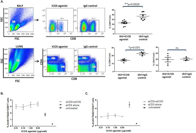
Figure 2. ICOS agonist treatment reduces the frequency of CD8+ T cells but does not affect the polyclonal CD4+ T cell compartment.
IAV infected BALB/c mice were treated with ICOS agonist or hamster IgG isotype (control antibodies) as depicted in Figure 1A. On day 7–9 post infection the percentage of CD8+ in BAL (day 7, pooled data from two independent experiments) and lung (day 8) as well as CD4+ T cells in lung (day 9) was determined by flow cytometry. The dots in all experiments represent data from individual mice (A). In vitro apoptosis assays were performed, as described in materials and methods, followed by annexin-V staining on live T cells and subsequent flow cytometric analysis. Dot plot graphs indicate percentages of apoptotic CD8+ and CD4+ T cells (mean of triplicate wells) plotted against increasing concentration of ICOS agonist antibody added to the culture. CD3 alone (mean of triplicate wells) represents that cells were stimulated by anti-CD3 treatment, in the absence of ICOS agonist; untreated (mean of duplicate wells) represents that cells were neither anti-CD3 stimulated nor treated with ICOS agonist. Column statistics were performed for the apoptosis assay (the data is expressed by mean/SEM) (B and C).

