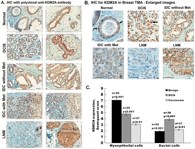
Figure 1. Myoepithelial cells of the breast express KDM2A.
(A) Immunohistochemical staining of KDM2A on human breast cancer tissue microarray. Immunostaining was performed using rabbit anti-human KDM2A antibody and representative images of KDM2A expression in normal breast, Ductal Carcinoma in situ, Infiltrating Ductal Carcinoma with and without metastasis, lymph node metastasis are shown. Magnification is 200X, scale bar = 50 µm. (B) Enlarged images of normal and cancer breast tissue sections. Arrows indicate positively stained myoepithelial cells. Scale bar = 50 µm. (C) Quantitative analysis of KDM2A in breast tissue microarray. The Immunostaining of KDM2A was quantified by using semi quantitative scoring method based on cellularity and intensity of expression. The means of two independent arrays are shown. All p-values were calculated using a two-sided Student t-test.

