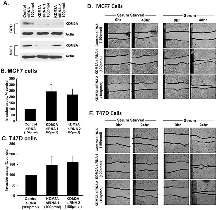
Figure 2. KDM2A suppresses invasion and migration of breast cancer cells.
(A) Western blot analysis showing decreased KDM2A levels in T47D and MCF7 cells with two different siRNAs to KDM2A (Ambion and Santa Cruz) compared to control siRNA. (B) Silencing KDM2A by KDM2A siRNA 1 increased invasion in MCF7 cells by 145±23% (p<0.05) and KDM2A siRNA 2 increased invasion by 118±21% (p<0.05) when compared to control siRNA. (C) In T47D cells, KDM2A siRNA 1 enhanced invasion by 48±28% (p<0.1) and KDM2A siRNA 2 by 64±16% (p<0.05) when compared to control siRNA. (D) Both KDM2A siRNA1 (100 pmol) and KDM2A siRNA2 (100 pmol) transfected MCF7 cells migrated into the wound in the presence of serum at 48 hr when compared to control siRNA transfected cells. Serum starved cells did not migrate into the wound with or without KDM2A suppression. (E) T47D cells additionally show significant migration into the wound after transfection with KDM2A siRNA1 and KDM2A siRNA 2 in the presence of serum at 24 hr when compared to control siRNA. Serum starved cells did not migrate into the wound with or without KDM2A suppression. Magnification-200X.

