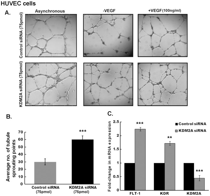
Figure 3. Silencing KDM2A enhances angiogenic tubulogenesis.
(A) HUVEC cells were transfected with either control siRNA or KDM2A siRNA (75 pmol). 24 hr later, cells were plated on matrigel in complete media (asynchronous) or with or without 100 ng/ml VEGF. Images were captured 18 hr later using Leica inverted microscope and representative images are shown. (B) Tubule sprouting points were estimated using Image Pro software. Ablation of KDM2A showed significant increase (2-fold, p<0.01) in the number of sprouting points. (C) Real-Time showing increased mRNA expression of FLT-1 (2.2±0.11-fold, p<0.01) and KDR (1.7±0.9-fold, p<0.05) receptors upon silencing KDM2A expression. Simultaneous decrease in KDM2A mRNA levels (p<0.01) is seen with 75 pmol of KDM2A siRNA compared to control siRNA.

