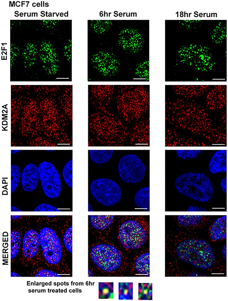
Figure 4. KDM2A co-localizes with E2F1 at 6 hr of serum stimulation.
Quiescent MCF-7 cells were serum starved for 48 hr and serum stimulated for 6 hr and 18 hr. Cells were fixed, permeabilized for 5 min with 0.2% Triton X-100/PBS and immunostained for E2F1 (anti-mouse IgG, green) and KDM2A (anti-rabbit IgG, red). Cells were visualized by confocal microscopy. E2F1 was predominantly localized in the nucleus (upper panels), while KDM2A was more ubiquitously distributed in the cells (middle panels). Co-localization of E2F1 and KDM2A was observed in the nucleus at 6 hr of serum stimulation and disappeared totally by 18 hr (right panels). Images were captured at 630X oil using DM16000 inverted Leica TCS SP5 tandem scanning confocal microscope. Scale bar = 200 µm. Pearson's correlation for co-localization at 6 hr was 1.0.

