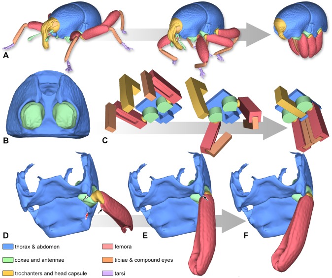
Figure 3. Blocking mechanisms of legs in Trigonopterus vandekampi.
(A) Illustration of the movement from walking position to thanatosis. (B) Prothorax in ventral aspect; note the flattened mesial faces of the coxae and the narrow thoracic canal. (C) Simplified model of the prothoracic blocking mechanism. (D–F) Metacoxal leverage. (D) Hind leg elevated; note the depressed face of the metafemur (black arrow), the metathoracic intercoxal ridge (white arrow) and the abdominal protrusion (red arrow). (E) Inward rotation of the trochanter causes the depressed face of the femur to press against the posterior face of the intercoxal ridge (arrow). (F) The leverage effect causes the coxa to swing backwards and the joint comes to a dead stop.

