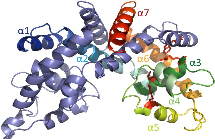
Figure 3.

Predicted model of MftR. MftR model based on the structure of HucR (2fbk), created using SwissModel in automated mode. One monomer is colored blue to red (amino-terminus to carboxy-terminus; helices are shown as α1 to α7) and the other is in purple. Conserved residues, which are predicted to bind urate, are in red stick representation.
