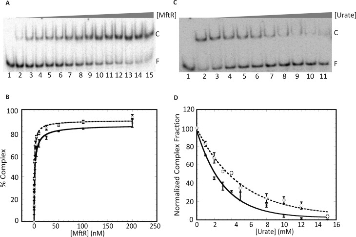
Figure 5.
MftR binds both palindromes in its operator DNA, and the complexes are sensitive to urate. (A) EMSA showing mftpO (3.0 nM) titrated with increasing concentrations of MftR (0.1–200 nM; lanes 2–15); reaction in lane 1 contains DNA only. Complex and free DNA are identified at the right as C and F, respectively. (B) Fractional complex formation plotted as a function of MftR concentration. Binding isotherm with mftrO (○; solid line) and mftpO (+; dashed line). (C) Effect of urate on the binding of MftR to mftpO. Lane 1 contains DNA only. Reaction in lane 2 contains no ligand. The MftR-mftpO complex was titrated with increasing concentrations of urate (3–18 mM; lanes 3–11). (D) Normalized complex fraction as a function of urate concentration. MftR-mftrO complex (○; solid line) and MftR-mftpO complex (+; dashed line) titrated with increasing concentrations of urate. Error bars represent the standard deviation of three independent repeats.

