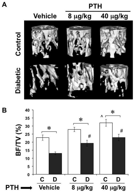
Figure 1. PTH treatment counteracted trabecular bone loss from T1-diabetes.
Diabetes was induced with STZ at 14 weeks of age. At the same time, control (citrate buffer only) and diabetic mice were started on a daily regimen of subcutaneous injections of PTH (8 or 40 μg/kg) or saline vehicle. PTH treatment was continued for the remainder of the study. Mice were harvested at 40 days after the first streptozotocin injection and tibias were analyzed by μCT. (A) Representative three-dimensional isosurface images of the trabecular bone of the proximal tibia of control and diabetic, vehicle and PTH treated mice. (B) BV/TV of control (white bars) and diabetic (gray bars), vehicle and PTH treated mouse tibias. Bars represent mean ± standard error. n ≥ 12 per group. Significance between groups was determined with a post-hoc test (only after factorial ANOVA determined significance). *p < 0.05 compared to treatment (vehicle, 8 or 40 μg/kg PTH) matched control. ^p < 0.05 compared to vehicle-treated control. #p < 0.05 compared to vehicle-treated diabetic.

