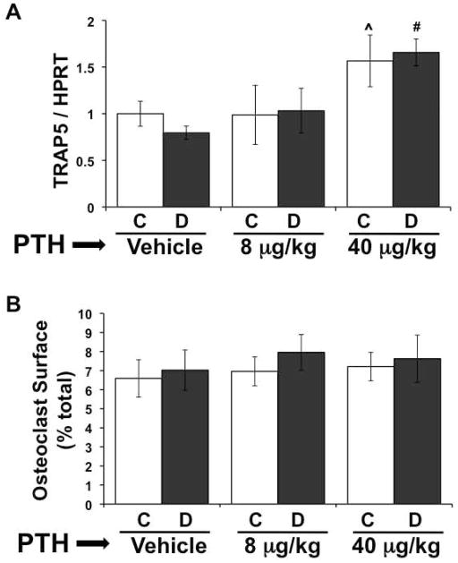
Figure 3. High dose PTH promotes bone resorption in diabetic mice.
A) mRNA from frozen tibiae was converted to cDNA and amplified with primers specific for tartrate resistant acid phosphatase (TRAP5) and the housekeeping gene HPRT. (B) Osteoclasts were identified with TRAP5 staining in the distal femur. Length of trabecular bone surface covered by osteoclasts was measured and expressed as a percent of the total bone surface. Bars represent mean ± standard error of control (C, white bars) and diabetic (D, gray bars), vehicle, 8 μg/kg and 40 μg/kg PTH treated mice at 40 days. PTH treatment was daily from 0 to 40 days. N = 4–8 per group. Significance between groups was determined with a post-hoc test (only after factorial ANOVA determined significance). *p < 0.05 compared to treatment (vehicle, 8 or 40 μg/kg PTH) matched control. ^p < 0.05 compared to vehicle-treated control. #p < 0.05 compared to vehicle-treated diabetic.

