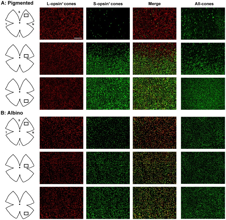
Figure 2. Opsin expression in pigmented and albino mice.
Magnifications from pigmented (A) and albino (B) mice flat mounted retinas showing L-opsin+cones, S-opsin+cones and their merged image. The rightmost column shows all-cones (both opsins developed with the same fluorophore). In the retinal drawings on the left, is shown the area where the magnifications were taken from. S-opsin+cones are sparse in the superior retina of the pigmented strain and abundant in the albino one. In both strains most genuine S-cones are found in the inferior retina. In the pigmented strain majority of genuine L-cones lay in the superior retina, while in the albino mouse majority of L-cones are dual. And so dual cones are found across the retina in the albino strain and mostly restricted to the ventral retina in the pigmented one. S: superior, N: nasal, I: inferior, T: temporal. Bar: 100 µm.

