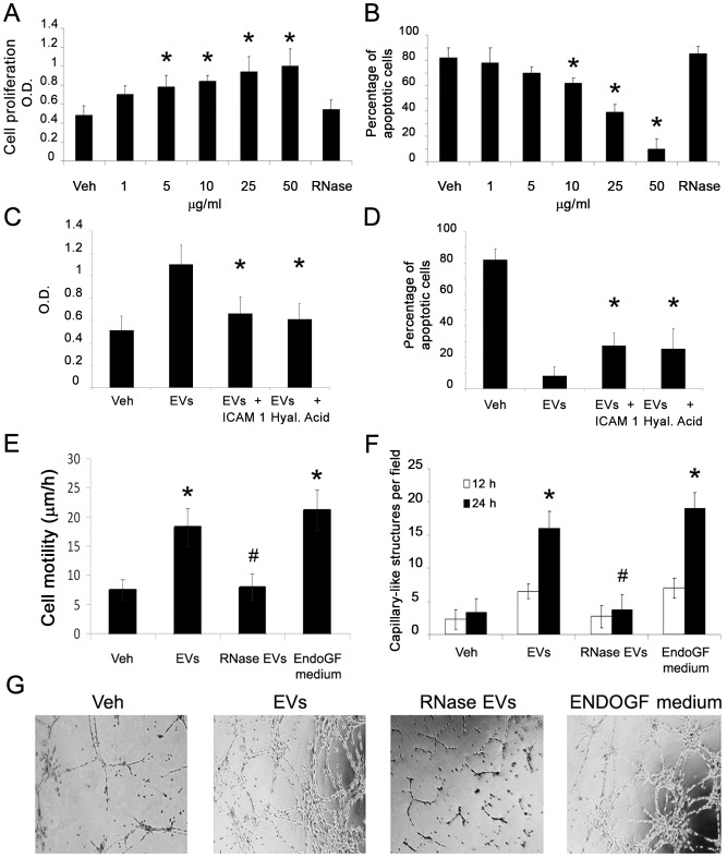
Figure 3. Pro-angiogenic and anti-apoptotic effects of EVs on IECs.
A) BrdU proliferation assay of IECs incubated for 12 hrs with different doses of EVs or with RNAse pre-treated EVs (*p<0.05 EVs vs. vehicle alone or RNase EVs). B) Apoptosis (TUNEL assay) of IECs incubated for 48 hrs with different doses of EVs or with RNAse pre-treated EVs (*p<0.05 EVs vs. vehicle alone or RNase pre-treated EVs). C) BrdU proliferation assay of IECs incubated for 12 hrs with EVs (50 µg/ml) pre-treated or not with a blocking antibody directed to ICAM-1 or with HA (*p<0.05 EVs+blocking monoclonal antibody or HA versus EVs alone). D) Apoptosis (TUNEL assay) of IECs incubated for 48 hrs with EVs (50 µg/ml) pre-treated or not with a blocking antibody directed to ICAM-1 or with HA (*p<0.05 EVs+blocking monoclonal antibody or HA versus EVs alone). E) Time-lapse videomicroscopy analysis of 6 hrs IEC migration induced by EVs (50 µg/ml), EVs pre-treated with 1 U/ml RNase or by medium enriched for endothelial growth factors (EndoGF medium) (*p<0.05 EVs or EndoGF medium vs. vehicle alone; #p<0.05 RNase EVs vs. EVs). F–G) count and representative micrographs of in vitro formation of capillary-like structures by IECs seeded on Matrigel-coated surfaces in the presence of vehicle alone, EVs (50 µg/ml), RNase pre-treated EVs or medium enriched for endothelial growth factors (EndoGF medium) for 12 hrs (white columns) or 24 hrs (black columns) (*p<0.05 EVs or EndoGF medium vs. vehicle alone; #p<0.05 RNase EVs vs. EVs).

