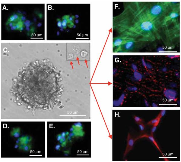
Figure 1. Multilineage Differentiating Stress Enduring (Muse) cell characterization by pluripotency markers and morphology.
Immunostaining indicates that Muse cells express the pluripotency markers: (A) stage-specific embryonic antigen-3, (B) OCT3/4, (D) SOX2 and (E) Nanog. (C) Muse cells derived from adipose tissue (Muse-AT) cells grow in suspension, forming cell clusters as well as individual cells (red arrows). (F) Muse-AT cells were grown as adherent cells in the presence of myocyte differentiation medium. Formation of myocytes was detected using an antihuman MSA antibody. Nuclei were stained with DAPI (blue). (G) Muse-AT cells were grown as adherent cells in the presence of hepatocyte differentiation medium. Formation of hepatocytes was detected using an antihuman a-fetoprotein antibody. Nuclei were stained with DAPI (blue). (H) Isolated Muse-AT cells were grown for 7 days as nonadherent cells and then cultured for an additional 7 days as adherent cells. Neural-like cells were detected by immunofluorescence using an antihuman MAP2 antibody. Nuclei were stained with DAPI (blue). (Original magnification was 600× for figures F – H).
Reproduced from [18].

