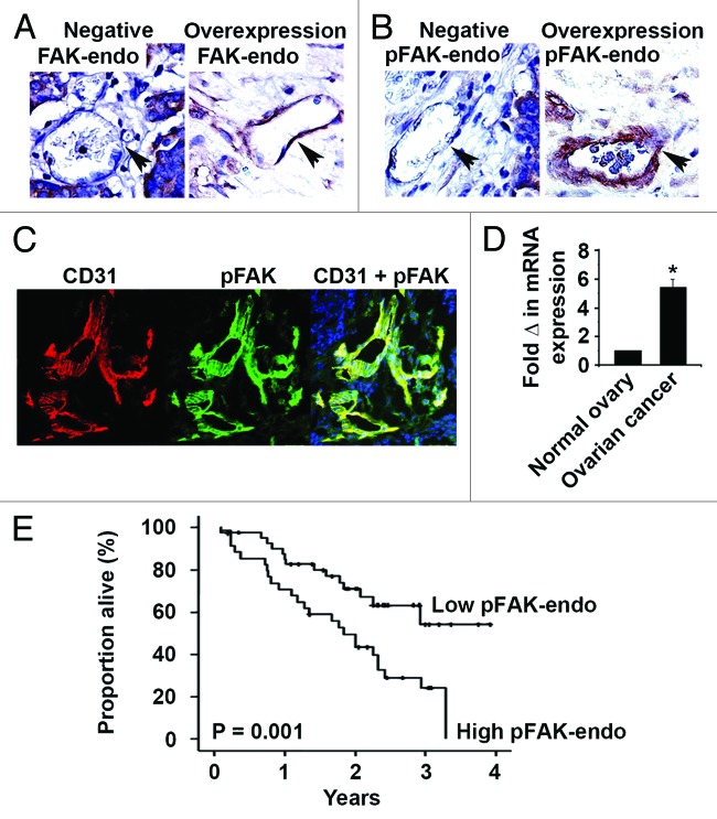
Figure 2. Tumor-associated endothelial cell FAK and pFAKY397 expression in human epithelial ovarian cancer. (A) Representative tumor-associated endothelial cell FAK (FAK-endo) immunohistochemical staining in advanced stage, high grade serous ovarian cancer specimens. Negative FAK-endo expression and FAK-endo overexpression are represented in the left and right panels, respectively. (B) Representative tumor-associated endothelial cell pFAKY397 (pFAK-endo) immunohistochemical staining in advanced stage, high grade serous ovarian cancer specimens. Negative pFAK-endo expression and pFAK-endo overexpression are represented in the left and right panels, respectively. Images were captured at original magnification 200×. (C) Representative CD31 (left panel) and pFAKY397 (center panel) immunofluorescent staining in advanced stage, high grade serous ovarian cancer. Dual CD31 and pFAKY397 staining (right panel) demonstrates activated FAK in the tumor vasculature. Images were captured at original magnification 200×. (D) Quantitative real-time PCR of endothelial cell FAK expression. FAK gene expression in 10 ovarian cancer isolates was calculated as the mean fold change relative to 7 normal ovarian endothelial specimens (normal = 1) using the 2−ΔΔCT method. (E) Kaplan–Meier plot depicting the impact of tumor-associated endothelial cell pFAKY397 (pFAK-endo) expression on ovarian cancer patient survival using the log-rank statistic.

An official website of the United States government
Here's how you know
Official websites use .gov
A
.gov website belongs to an official
government organization in the United States.
Secure .gov websites use HTTPS
A lock (
) or https:// means you've safely
connected to the .gov website. Share sensitive
information only on official, secure websites.
