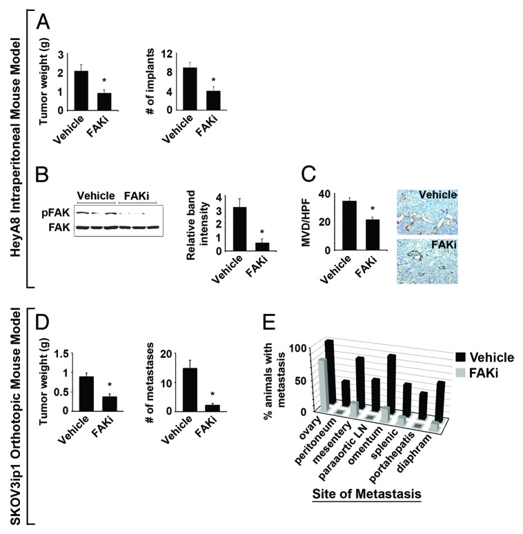
Figure 4. Effect of VS-6062 induced pFAKY397 inhibition on tumor growth and metastasis in HeyA8 intraperitoneal and SKOV3ip1 fully orthotopic mouse models of ovarian cancer. (A) Effect of VS-6062 (50mg/kg gavaged BID) on mean HeyA8 tumor weight and number of tumor implants ± SE. (B) Assessment of pFAKY397 expression by western blot to confirm target modulation in VS-6062 treated tumors. The histogram represents mean pFAKY397 relative to total FAK band intensity determined by densitometry ± SE. (C) Mean HeyA8 tumor microvessel density (MVD) evaluated by CD31 immunohistochemical staining (rat anti-mouse CD31 monoclonal antibody, DAKO) following treatment with VS-6062 or vehicle control ± SE. Representative photomicrographs appear above the histograms. Images were captured at original magnification 200×. (D) Effect of VS-6062 (50 mg/kg gavaged BID) on mean SKOV3ip1 tumor weight and number of metastases ± SE. (E) Effect of VS-6062 treatment on site-specific metastasis. The histogram displays the percent of animals with persistent ovarian involvement and metastasis to other organ sites following treatment with vehicle control vs. VS-6062. Paraaortic LN, paraaortic lymph nodes; FAKi, FAK inhibitor VS-6062; Vehicle, AEE (sterilized 90% polyethylene glycol 300 and 10% 1-Methyl-2-pyrrolidinone) diluent alone was used for all in vivo experiments.

An official website of the United States government
Here's how you know
Official websites use .gov
A
.gov website belongs to an official
government organization in the United States.
Secure .gov websites use HTTPS
A lock (
) or https:// means you've safely
connected to the .gov website. Share sensitive
information only on official, secure websites.
