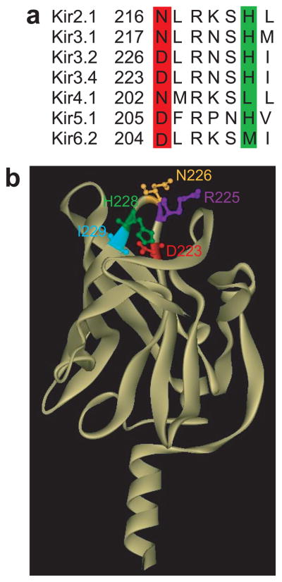
Figure 1.

Positions of key residues in a loop of Kir channels where Na+ is coordinated. (a) Alignment of the loop segment of Kir2.1, Kir3.1, Kir3.2, and Kir3.4, Kir4.1, Kir5.1 and Kir6.2, which includes the residues found in this study to coordinate the Na+ ion (Asp in the red highlighted position and His in the green highlighted position). (b) Cytosolic domain of the homology model of Kir3.4 based on the crystallographic structure of the cytosolic domain of Kir3.1 showing along with the three residues of focus in the present study (D223 -in red, H228 -in green, R225 -in purple) the neighboring asparagine N226 (yellow), and isoleucine I229 (cyan) residues.
