Figure 3.
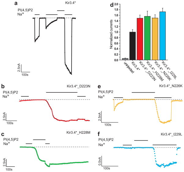
Experimental evidence for Kir3.4* residues involved in Na+ sensitivity (a)–(c) Representative traces of inside-out macropatch recordings of Xenopus oocytes. Na+ (30mM) and PIP2 (2.5μM) were applied as indicated by the bars in the control solution (ND96K+EGTA) (a) Kir3.4* (b) Kir3.4*_D223N (c) Kir3.4*_H228M. (d) Whole-cell basal currents of Kir3.4*, Kir3.4*_D223N, and Kir3.4*_H228M recorded in Xenopus oocytes at −80mV. (e)–(f) Representative traces of inside-out macropatch recordings of Xenopus oocytes. Na+ (30mM) and PIP2 (2.5μM) were applied as indicated by the bars in the control solution (ND96K+EGTA). Both Kir3.4*_N226K (e) and Kir3.4*_I229L (f) show strong sensitivity to Na+ similar to Kir3.4*.

