Figure 5.
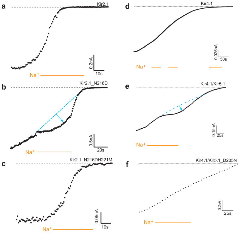
Experimental evidence for Kir2.1 and Kir5.1 residues involved in Na+ sensitivity (a)–(c) Representative traces of inside-out patches of Xenopus oocytes, showing Kir2.1 current rundown. Na+ (30mM) was perfused in the control solution (ND96K+EGTA). The rundown of Kir2.1_N216D is slowed down following application of sodium. This effect is reversed for the Kir2.1_N216DH221M mutant. (d)–(f) Representative traces of inside-out macropatch recordings of currents from Kir4.1 as a homomer or heteromer with Kir5.1expressed in Xenopus oocytes. Na+ (30mM) was applied as indicated by the bars in the control solution (ND96K+EGTA) The rundown of Kir4.1/Kir5.1 is slowed down following application of sodium. (d) Kir4.1 (e) Kir4.1/Kir5.1 (f) Kir4.1/Kir5.1_D205N

