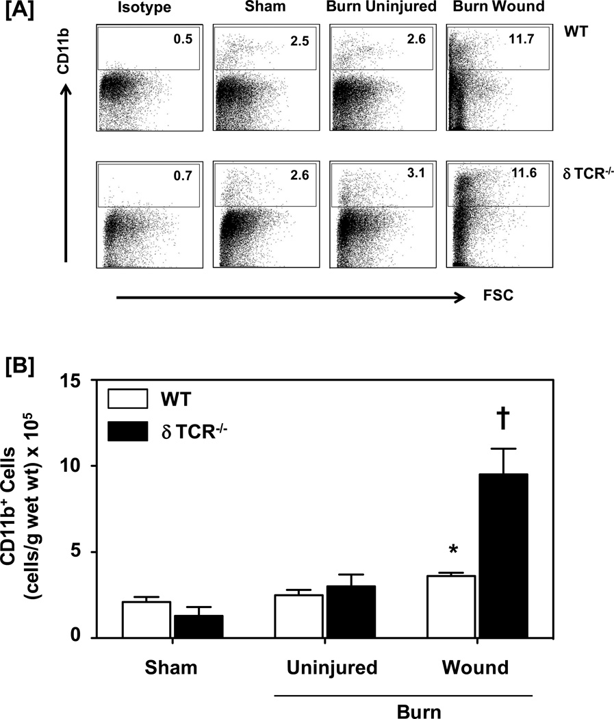
Figure 1.
Impact of γδ T-cells on wound CD11b+ myeloid cells. Three days after sham or burn procedure, skin cells from WT or δ TCR−/− mice were prepared and studied for CD11b+ myeloid cell characterization using flow cytometry. [A] Gating strategy. CD11b+ cells from the lymphocyte/myeloid cell gate of WT (Fig. 1A, Upper Panel) and δ TCR−/− (Fig. 1A, Lower Panel) mice. Representative dot plots are shown from sham, burn uninjured and burn injured skin cells. The numbers indicate the percentages of CD11b+ cells as determined by flow cytometry. [B] The number of CD11b+ cells as normalized to gram wet weight of the skin tissue. Data are mean ± SEM for 3–7 mice/group; * p<0.05 vs. uninjured skin of the respective WT or δ TCR−/− mice. † p<0.05 vs. burn wound of the respective WT mice.

