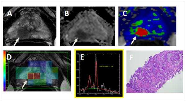
Figure 2.

A 63-year-old man with serum PSA of 3.28 ng/mL. (A) Axial T2W MRI, (B) ADC map of DW image, and (C) ktrans map derived from DCE MRI demonstrate a 1.3cm right apical mid-peripheral zone lesion (arrow). (D, E) MR spectroscopy was diagnostic and positive with a choline/citrate ratio of 0.623 +/− 13% (arrow). (F) Subsequent transrectal ultrasonography/MRI fusion-guided biopsy revealed Gleason 4+4 cancer in the lesion (up to 80% core involvement).
