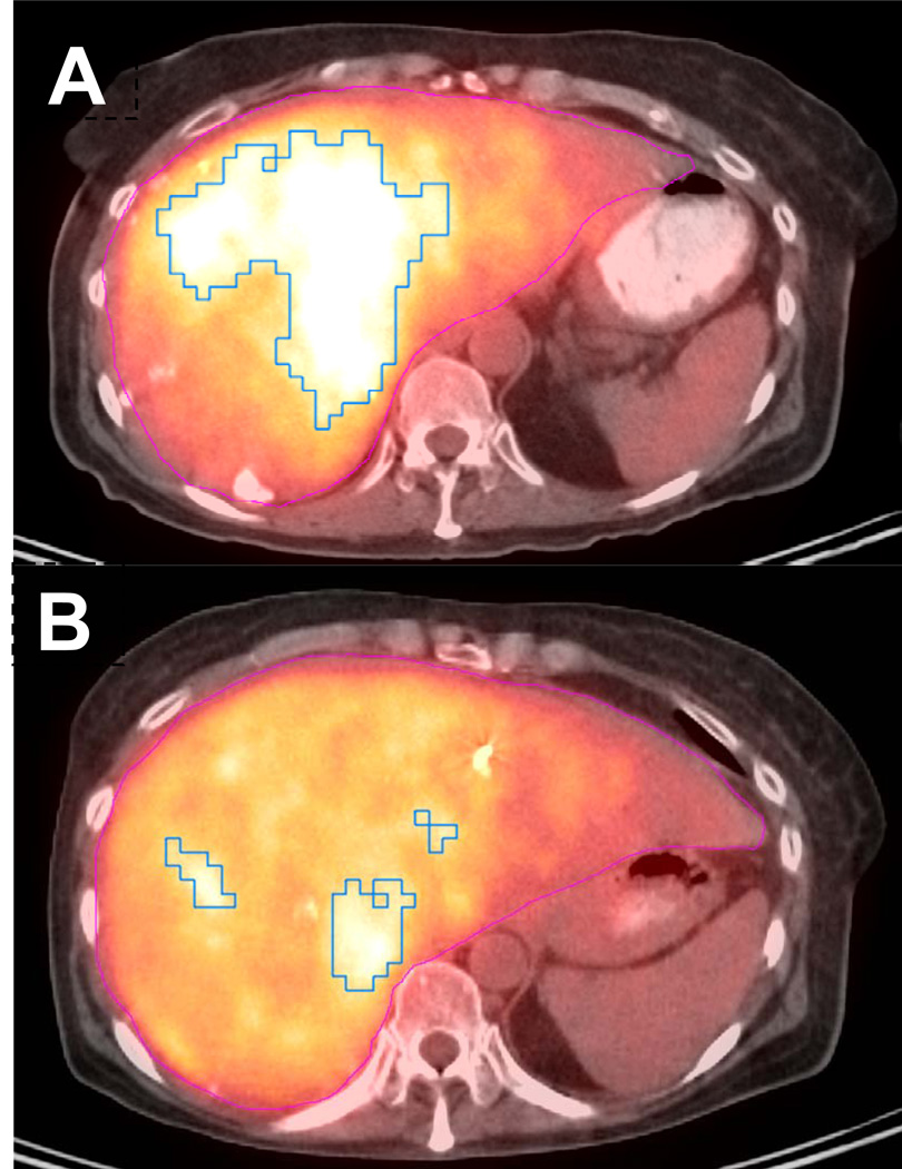
Figure 1.
Representative axial FDG PET/CT image from a patient with a partial response following 1.59 GBq of 90Y microspheres embolized via the left hepatic artery. (A) Pre-treatment functional tumor parameters improved from SUVmax 6.0, MTV 325 cc, and TGA 1667 cc to (B) SUVmax 5.2, MTV 117 cc, and TGA 551 cc on FDG PET/CT completed 88 days after treatment.

