
Figure 6. Oxidative stress increases DC transendothelial migration and CCR7 expression.
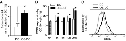
(A) Control DC and OS-DC (incubated for 24 h with 500 μM H2O2) were perfused under physiologic shear stress in a HUVEC-transwell flow chamber. A higher percentage of transendothlial migration was observed in OS-DC compared with controls (*P=0.026; n≥3 in each group). (B) DC were incubated in medium alone (DC) or in the presence of 500 μM H2O2 (OS-DC) for different time-points, and CCR7 expression on CD11c+ cells was assessed by flow cytomety. CCR7 expression was increased in OS-DC compared with control DC, with the highest expression observed at 24 h post-H2O2 treatment (*P=0.02 at 4 h, *P=0.07 at 6 h, *P=0.005 at 12 h, and *P<0.001 at 24 h, n=5 each group). (C) Representative histograms showing CCR7 expression on CD11c+ cells from control DC (gray curve) and 500 μM OS-DC for 24 h (black curve).