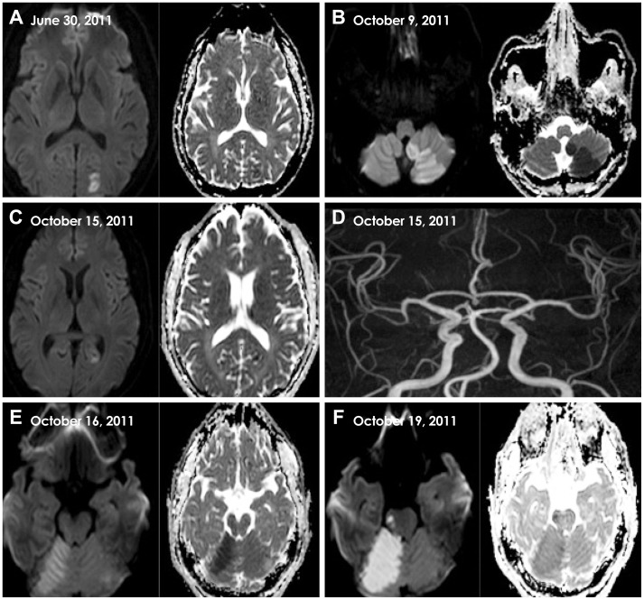
Fig. 1.
Serial MRI findings of five recurrent strokes in the present case. A: Three months previous to the current admission, DWI and ADC revealed acute left PCA infarction. B: On day 0, MRI revealed acute left PICA infarction. C: On day 6, the patient had a new ischemic infarction in the left PCA territory. D: MRA revealed normal intracranial arteries including in the vertebrobasilar system. E: On day 7, MRI revealed a new ischemic infarction in the right superior cerebellar artery territory. F: On day 10, MRI demonstrated another new ischemic infarction, this time in the right pons and involving the medial longitudinal fasciculus. ADC: apparent diffusion coefficient, DWI: diffusion-weighted imaging, MRA: magnetic resonance angiography, MRI: magnetic resonance imaging, PCA: posterior cerebral artery, PICA: posterior inferior cerebellar artery.

