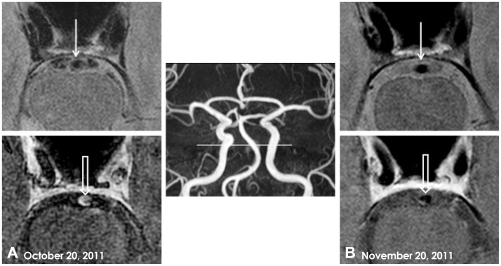
Fig. 2.
Intracranial arterial wall imaging using high-resolution, 3-tesla, contrast-enhanced MRI. A: Axial PD image (upper) showing eccentric vessel-wall thickness (closed arrow), and a three-dimensional postcontrast T1-weighted image (lower) demonstrating concentric enhancement of the proximal BA wall (open arrow). B: On day 31, a repeat axial PD image (upper) and postcontrast T1-weighted image (lower) obtained after steroid treatment showed decreases in both wall thickness (closed arrow) and wall enhancement of the proximal BA (open arrow). BA: basilar artery, MRI: magnetic resonance imaging, PD: proton-density-weighted.

