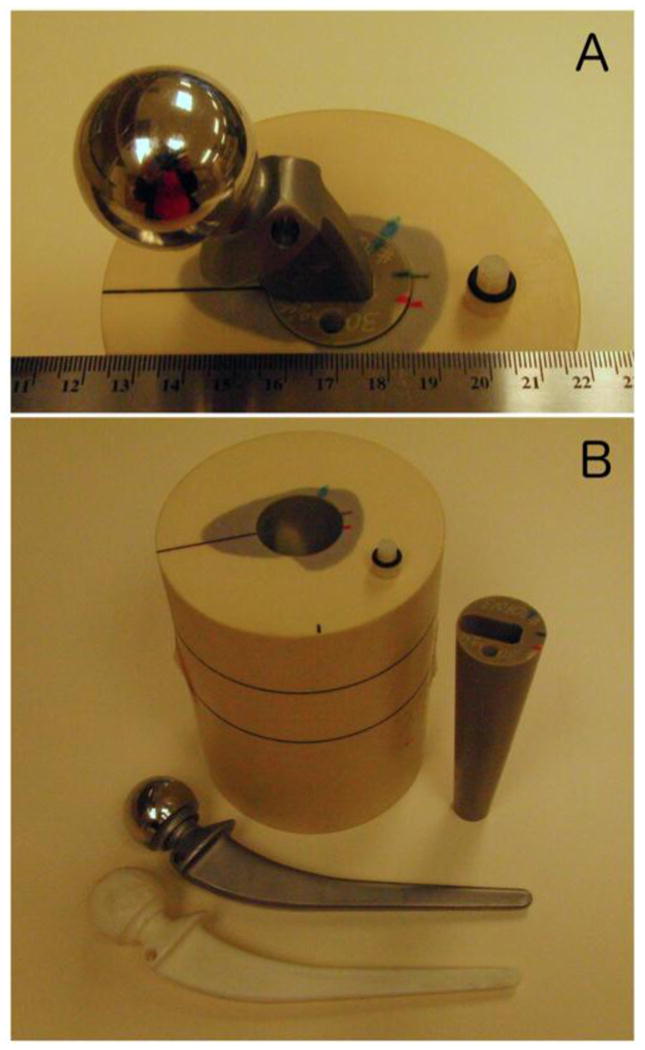
Figure 1.

Photographs of the hip phantom. In (A) the metal prosthesis is inserted into the bone mimicking cone, which is inserted into the simulated proximal femur embedded in the Plastic Water cylindrical body of the phantom. The 5 mm diameter cavity where activity was injected is visible toward the bottom of (A). In (B) the phantom has been disassembled into its constituent components; Plastic Water cylinder with simulated proximal femur inlay, conical bone mimicking insert with cylindrical cavity and pressfit-prosthesis bore, metal prosthesis, and plastic prosthesis replica.
